
Eye Conditions and Disorders
We are proud to bring you the very best clinical eyecare available. We diagnose and treat all kinds of eye health issues and take the time to explain any eye conditions you may have, as well as treatment options available to you.
Amblyopia is a neuro-developmental condition in which vision does not develop adequately in one eye. Amblyopia may be caused by any condition that affects normal visual development or use of the eyes including an eye turn, uncorrected refractive error, or ocular pathology which blocks light from reaching the retina such as a cataract. In each of these cases, the brain chooses to ignore the image produced by the affected eye (known as suppression), which inhibits the growth and development of the nerve fibers of the affected eye. Most children adapt well to seeing through one eye, so many parents are unaware that the child has a problem and the condition often goes undiagnosed. All children are recommended to have a thorough eye examination before they start school.
Astigmatism is a condition in which the shape of the cornea – the clear window at the front of the eye – is unequal in different directions. A useful analogy is to compare the shape of a soccer ball with a rugby or AFL ball. A soccer ball is spherical in shape with equal curvatures in all meridians. The surface of a rugby or AFL ball on the other hand, has different curvatures in the horizontal and vertical meridians i.e. the horizontal meridian is flatter, whilst the vertical meridian is steeper. The shape of a normal cornea typically resembles the shape of a soccer ball whereas an astigmatic cornea can be likened to the shape of a rugby or AFL ball. Symptoms of astigmatism include headaches, fatigue, eyestrain, as well as distorted or blurred vision.
Blepharitis is a common condition that causes inflammation of the eyelids. Blepharitis is often associated with overgrowth of bacteria or a parasite known as demodex on the eyelashes or eyelid margins. Blepharitis can affect people of all ages and is often associated with certain skin conditions including acne, rosacea and seborrheic dermatitis. Common signs and symptoms of blepharitis include chronic irritation, redness of the eyelid margins and crusting of the eyelashes (particularly in the morning).
Cataract is a condition which causes clouding of the clear lens in the eye and is one of the leading causes of vision impairment. Cataracts can be congenital yet more commonly occur with age as the focusing lens in the eye becomes progressively less clear, disrupting the flow of light through the eye and onto the retina. Cataracts are generally slowly progressive but certain types of cataracts can progress more quickly. Common symptoms of cataract include blurred vision, sensitivity to glare and distorted or double vision.
Colour vision deficiencies (CVD) (sometimes called colour blindness) represent a group of conditions that affect the perception of colour. Red-green colour vision defects are the most common form of colour vision deficiency.
CVDs are almost always inherited in an X-linked recessive pattern, although they can be acquired as a result of particular diseases and injuries. Males are affected by X-linked recessive disorders much more frequently than females since males have only one X chromosome and females have two, both X chromosomes on a female would have to have a genetic change in order to cause the disorder.
Males inherit their X chromosome from their mother and their Y chromosome from their father. Therefore, if a mother is a carrier of a CVD on one of her two X chromosomes, her son has a 50/50 chance of inheriting a CVD. Since females have two X chromosomes, one from the mother and one from the father they have less of a chance of being affected. About 8% (1 in 12) of males and 0.5% (1 in 200) of females have CVDs.
CVDs affect the ability to discriminate between certain colours such as red and green. A person with a CVD will try to discriminate colour differences by using other cues such as brightness differences. CVDs often do not impair quality of life but can affect career choices where colour discrimination is important for safety reasons.
Diabetic retinopathy is a complication of diabetes where the tiny blood vessels in the retina at the back of the eye become damaged and begin to leak blood and other fluids, which can lead to vision loss.
Most people, who have suffered from diabetes for over 20 years, will have some degree of retinopathy (nearly all patients with Type 1 diabetes and 58% of patients with Type 2 diabetes).
Early signs of diabetic retinopathy include small haemorrhages and microaneurysms appearing in the retina. In later stages, excessive microvascular leakage can cause swelling at the macular (the region of the retina responsible for all detailed central vision).
Symptoms of diabetic retinopathy include blur or loss of vision. For diabetics, strict control of blood sugar levels is essential in preventing the progression of diabetic retinopathy.
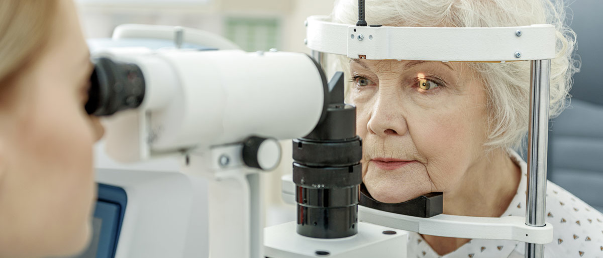
At Vedelago Optometrists we utilise Digital Retinal Imaging (DRI) and Optical Coherence Tomography (OCT) to detect diabetic retinopathy. Digital retinal imaging photographs the surface of the retina looking for signs of haemorrhages, lipid exudates or micro-aneurysms. OCT is a non-invasive imaging test, which uses light waves to take cross-sectional images of the retina. OCT allows the optometrist to see each of the retina’s distinctive layers allowing them to detect leaking blood or fluid beneath the surface of the retina.
i Hietala K, Harjutsalo V, Forsblom C, Summanen P, Groop PH; FinnDiane Study Group. Age at onset and the risk of proliferative retinopathy in type 1 diabetes. Diabetes Care. 2010;33(6):1315–1319. doi:10.2337/dc09-2278
Dry eye is a common condition, which affects one in four people worldwide and is more likely to occur in women and the elderly. Symptoms of dry eye include burning, scratching, irritation, redness and watering of the eye, as well as blurred vision. Although dry eye is generally not a sight-threatening condition, it can cause significant discomfort for sufferers. Dry eye can be caused by insufficient tear production from the lacrimal gland or an unstable lipid layer, which is the thin oily layer on the outer most part of the tear film.
Floaters are deposits of natural materials that are present within the eye’s vitreous humour. The vitreous humour is a clear jelly located inside the eye that is attached to the retina. Floaters typically increase in number as we age, however they may also be caused by disease or injury. Floaters are visible because of the shadows they cast on the retina. Common symptoms of floaters include the appearance of spots, threads, fragments or cobwebs, floating slowly in the visual field.
Floaters are generally harmless yet can be associated with retinal holes or tears which can increase the risk of developing a retinal detachment. Anyone who notices the sudden onset of floaters is recommended to have a Dilated Fundus Examination (DFE) performed where eye drops are instilled in the eye to dilate (expand) the pupil. A dilation allows the optometrist to see to the very edge of the retina where retinal holes and tears commonly develop.
Glaucoma is an eye condition which is characterised by damage to the optic nerve and retina, causing progressive vision loss. Glaucoma is often, but not always, associated with increased pressure of the fluid in the eye (aqueous humour). Glaucoma has few to no symptoms in the early stages; it is not until the condition is more advanced does the sufferer become aware of vision loss. At Vedelago Optometrists we utilise Optical Coherence Tomography (OCT), a non-invasive imaging test to detect glaucoma. OCT uses light waves to take cross-sectional images of your retina. OCT allows the optometrist to see each of the retina’s distinctive layers, which can be used to map and measure their thickness. The retinal nerve fibre layer thickness measurements help with the diagnosis of glaucoma. Studies have shown that OCT technology can detect glaucomatous damage up to 8 years before vision loss.
i Kuang TM et al. Ophthalmology 2015. Oct;122(10):2002-9.doi:10.1016/j.ophtha.2015.06.015. Epub 2015 Jul
Hyperopia, also known as long-sightedness, refers to a refractive condition where the focusing power of the eyeball is too weak in relation to the length of the eye. Hyperopia develops as a result of the visual image being focused behind the retina rather than directly on it.
Hyperopic people must exert extra effort to bring their near vision into sharp, clear focus. Symptoms may include; blurriness when reading, eyestrain, fatigue and or headaches after sustained close work and difficulty adjusting focus. Higher degrees of hyperopia can also affect distance vision especially with increasing age.
Keratoconus is a condition which causes the cornea, the clear window at the front of the eye to become progressively thinner. As a result of this thinning, the normally round shape of the cornea becomes distorted and a cone-like bulge develops, resulting in significant visual impairment. A useful analogy is to compare the shapes of an orange with a pear. The average person has a spherical shaped cornea like an orange whereas a person with keratoconus has a bulge, generally in the lower region of the cornea much like a pear. Keratoconus is typically diagnosed in the patient’s adolescent years and may progress until the patient is in their twenties and thirties.
We utilise corneal topography and corneal pachymetry technology to measure the curvature and thickness of the cornea respectively, both of which help confirm the diagnosis of keratoconus. Patients with keratoconus may require rigid gas permeable contact lenses to enhance their vision and those with more advanced forms may require surgery.
Macular degeneration is a condition that causes progressive macular damage resulting in loss of central vision. Macular degeneration is the leading cause of blindness in Australia with 1 in 7 people over the age of 50 affected to some degree, while incidences increase with age*.
The macular is the central part of the retina, the light sensitive tissue at the back of the eye which processes all central visual images. It enables us to read, recognise faces, drive safely and see colours clearly.
Common symptoms of macular degeneration include distorted or blurred vision, the need to use greater amounts of light to read and central vision loss.
We utilise Optical Coherence Tomography (OCT), a non-invasive imaging test to detect macular degeneration. OCT uses light waves to take cross-sectional images of your retina. OCT allows the optometrist to see each of the retina’s distinctive layers and pick up early signs of macular degeneration, which can include fatty deposits known as drusen, pigment cell disruption or leaking blood or fluid.
*Source: Macular Disease Foundation.
Myopia is also known as short-sightedness and is a refractive condition, which causes blurred distance vision. Myopia develops as a result of the visual image being focused in front of the retina rather than directly on it. Myopia is caused by the eyeball being too long or the focusing power of the eye being too strong.
Symptoms of myopia include squinting, as well as blurred distance vision. This may be more obvious when viewing the television or trying to read signs while driving.
While the exact cause of myopia is unknown, it is believed to be a combination of nature (genetics) and nurture (environment). Repeated near work i.e. reading, computer and device use has been shown to increase the risk of developing myopia.
Presbyopia is a condition that impairs the ability to focus on objects up close. Symptoms of presbyopia are usually noticed in one’s forties and fifties and include eyestrain, difficulty seeing in dim light and problems focusing on small objects and/or fine print. Presbyopia is a normal ageing condition that develops due to hardening of the crystalline lens inside the eye, which is responsible for focusing on near objects.
Pterygium is a wing-shaped growth of vascular tissue which occurs commonly on the nasal side of the conjunctiva, the white part of the eye. Pterygium is most commonly caused by ultraviolet (UV) damage but can be exacerbated by windy and dusty conditions. While many pterygia remain stable over time, some grow onto the cornea (the clear window at the front of the eye) resulting in blurred or distorted vision.
Pterygia can be prevented by wearing sunglasses or protective eyewear particularly in the first two decades of life.