The Run-down on Glaucoma
Glaucoma is the name given to a group of eye diseases where vision is lost due to damage to the optic nerve. Glaucoma is known as the ‘thief of sight’ because the loss of sight occurs gradually and a considerable amount of peripheral (side) vision is lost before many people are aware they even have a problem. Unfortunately, there is no cure for glaucoma, and vision loss is irreversible, but if detected early, treatment is able to slow down and, in many cases, even stop the progression of the condition. The key is early identification and the best way to do this is by having regular eye exams.
What are the types of glaucoma?
There are up to 40 types of glaucoma, which can be subdivided into primary and secondary glaucoma.
Primary glaucoma is a subset of glaucoma defined by an open, normal appearing anterior chamber angle and raised intraocular pressure (IOP), with no other underlying disease. If there is an identifiable underlying cause for raised IOP, i.e., injury, surgery or other eye disease, this is termed secondary glaucoma.
In healthy eyes, the rate of fluid production equals the rate it flows out of the eye, which maintains a stable intraocular pressure. Fluid (otherwise known as aqueous humour) flows out of the eye through a spongy tissue known as the trabecular meshwork. The trabecular meshwork sits in the ‘drainage angle’ between the iris (the coloured part of the eye) and the cornea (the clear, protective outer layer). If the outward flow of fluid is reduced or blocked, a build-up occurs, and this leads to an increase in IOP. The higher the intraocular pressure, the more likely a person is to develop glaucoma. However, high IOP in itself is not sufficient to confirm a diagnosis of glaucoma. In fact, up to 30-40% of all people with glaucoma actually have pressure within the normal range, which is known as normal tension glaucoma.
What are the symptoms of glaucoma?
Glaucoma usually affects the peripheral (side) vision first and is generally not associated with any other symptoms so as a result many people can remain unaware they have the condition until their eyesight is significantly compromised. The exception is a condition known as angle closure glaucoma, a more severe but thankfully less common form of glaucoma which is characterised by the sudden closure of the drainage angle within the eye. It results in the complete blockage of the trabecular meshwork and a rapid increase in eye pressure that causes vision to deteriorate quickly. Angle-closure glaucoma is considered a medical emergency that requires urgent treatment to prevent sight loss. People with angle closure glaucoma can get blurred vision, pain and a red eye, and might see haloes around bright lights. Yet, even in this condition there may be few symptoms present in the early stages.
What causes glaucoma?
Glaucoma occurs due to the pressure increasing in the eye, due to the gradual blockage of the trabecular meshwork and drainage angle. There is also a relationship between the pressure within the eye and the cerebro-spinal fluid pressure behind the optic nerve. If the intra-ocular pressure is too high and the cerebro-spinal fluid pressure too low damage occurs to the optic nerve, which is responsible for transmitting the visual signal to the brain.
Who is at risk of glaucoma?
You are at higher risk of developing glaucoma if you have:
- Family history of glaucoma
- High intra-ocular pressure (IOP)
- Aged over 50
- Short-sighted or long-sighted
- Use of cortisone (steroid) medications for long periods
- Diabetes
- High or low blood pressure
- Suffer migraine headaches
- History of an eye operation or eye injury
How is glaucoma diagnosed?
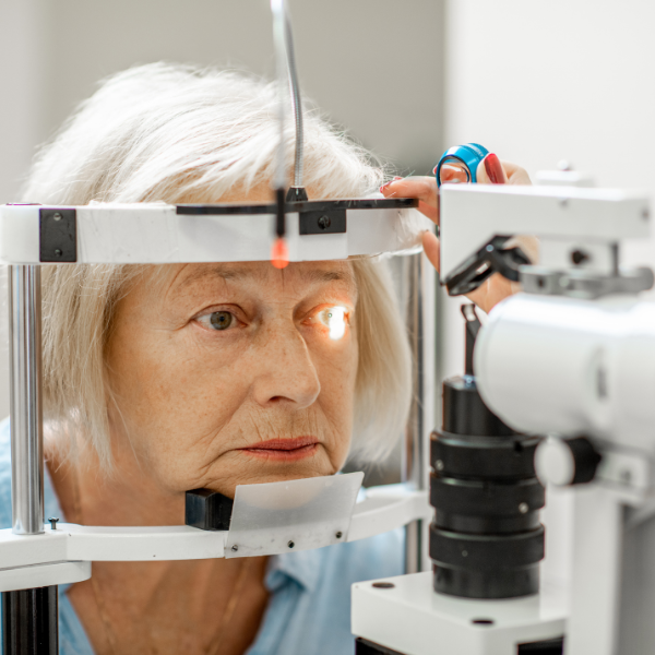
An optometrist is able to detect glaucoma as part of a thorough eye examination.
There are four major tests to detect glaucoma.
- Tonometry: A device is used to measures the fluid pressure within the eye.
- Optical Coherence Tomography (OCT): Is a computerised ultrasound that measures the thickness of the retinal nerve fibres. Glaucoma causes the gradual thinning of the retinal nerve fibre layer, which if becomes too pronounced causes a loss of vision. The OCT measures the thickness of the retinal nerve fibre layer to the micron or one-thousandth of a millimetre. In many cases an OCT is able to detect glaucoma before vision is lost.
- Visual fields test: Glaucoma causes blind spots to develop in the peripheral or side vision. A visual field analyser is a computerised instrument that is able to test the sensitivity of your peripheral vision.
- Gonioscopy: Evaluates the eyes drainage network to see whether there are any signs of blockage.
A thorough glaucoma examination will take approximately 30 to 45 minutes.
Treatment of Glaucoma
Glaucoma is treated by reducing the eye’s intra-ocular pressure. The primary way to do this is with eye drops. Glaucoma drops work by two primary mechanisms, one is to reduce the production of fluid within the eye, the other aims to improve the drainage of fluid out of the eye. In difficult to control cases often a combination of the two types of drops can be used.
The second way to lower pressure is with the use of laser, which is applied to the trabecular meshwork to facilitate improved drainage of fluid out of the eye. The laser is quick and painless and is becoming the treatment of choice since it may remove the need to use drops, which people can often forget to use and can also cause eye irritation.
The third option is to have a stent inserted in the eye’s drainage angle, which can help facilitate improved drainage of the aqueous humour. Micro-stents are usually performed at the same time as cataract surgery, which has also been shown to help reduce intra-ocular pressure.
If pressure cannot be controlled with medications and ongoing damage to the optic nerve occurs then glaucoma drainage surgery may be required. Drainage surgery involves surgically cutting the eye and inserting a drainage tube, it is a more invasive procedure but can reduce pressure more than other treatment options.
Summary
The key to effectively managing glaucoma is early identification. We recommend all people over the age of 50 have a glaucoma check once every 3 years, or sooner if you have any significant risk factors.
Please contact us to arrange a test today.

Diagnosing Dry Eye Syndrome
Our eyes are the most sensitive and easily disturbed part of your body. Even with slight irritation, they may start watering. Therefore, we have to take extra care in order to maintain eye health and prevent inflammation, especially with aging. It is therefore important to understand the symptoms of dry eye syndrome, and how it should be treated.
Tears & Why They’re Important
The eyes produce tears to remove irritants and to keep our eyes lubricated. Tears are made up of:
- Mucus
- Oil
- Antibodies
- Water
The above-quoted ingredients come from special glands around your eyes. An excess tear-flow from your eyes can occur due to poor tear drainage or overproduction of tears. Watery eyes are often not harmful but can be the cause of irritation. Alternatively, dry eye condition means that glands around the eyes aren’t working properly, and cannot adequately moisturise the eye.
What Happens If Tears Don’t Work Properly?
The production of tears is a natural cleaning mechanism, flushing away foreign objects that may come into contact with the eye easily. With the dry eye syndrome, the eye can not remove irritants effectively, and one of the two things can happen; insufficient or excessive production of tears. Inadequate production of tears may cause:
- Redness
- Irritation
- Continuous discharge of mucus
- Swelling
- Blurred vision
- Light sensitivity
If your eyes drops out water continuously, the result is often Reflex Tearing. This is because your eyes will send a distress signal to your nervous system to have the eyes lubricated to overcome the irritation and dryness. And as a result, excessive tear production will start.
Causes Of Dry Eye Syndrome?
There are many reasons described by science for this syndrome. However, the main ones are:
- An unbalanced tear flow system
- Dried tear film
- Drug-induced side effects
- Natural aging process
- Menopause
- Sjogren’s syndrome
- Rheumatoid arthritis
- Collagen vascular diseases
- Lagophthalmos
- Blepharitis
- Uneven eyelids
- Long-term use of contact lenses
How Is The Syndrome Diagnosed?
You need to go through a comprehensive eye examination to determine the exact quality and quantity of your tears that are produced. Furthermore, your doctor will go through some more procedures to determine the exact cause of the syndrome, and those tests will include:
- General medical history to note the symptoms and health problems
- Environmental causes and age factor that may add more to dry eye condition
- External assessment of the eye like the eyelid structure and blink dynamics
- Eyelid and cornea evaluation with bright light and magnifying glasses
- Tear abnormality along with quality and quantity difference
- Insertion of special dyes will be performed to observe the tear flow along with changes in the outer surface
Once your optometrist/doctor performs all of the above tests, then he/she will be suggesting the best treatment based on the current situation to smoothen out your dry eye condition.
Various Types Of Treatments For Dry Eyes
There are a lot of treatments available for treating dry eye syndrome. However, here are a few that your healthcare provider will prescribe depending on the severity of your condition. These are:
- Artificial ointments and teardrops
- Conserving tears
- Non-dissolving punctal plugs
- Punctal occlusion by cautery
- Lipiflow
- Temporary Punctal occlusion
- Cequa
- Testosterone cream
- Lifitegrast
- Xiidra
- Fish oil
These solutions are not to be administered without an expert optometrist’s prescription and advice. Visit us today to book your eye test consultation with our expert doctors and optometrist.

Holiday Trading Hours
Holiday Trading Hours
Our Practice will be closed from Friday, December 24 and will re-open on Tuesday, January 4.
On behalf of everyone at Vedelago, we hope you have a safe and happy holiday season.